Anatomy & Physiology
Scanning electron microscope (Focus Topic)
Summary
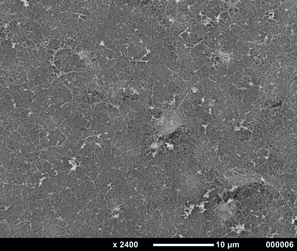
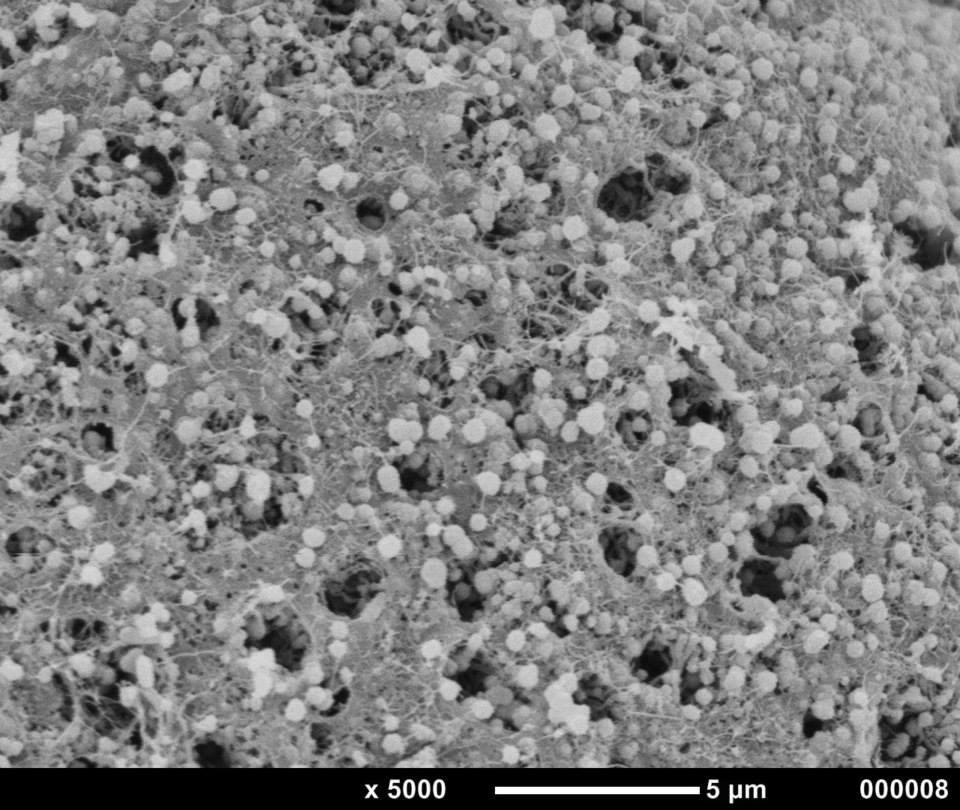
Samples of E. godeffroyi tentacles and skin were further examined under a scanning electron microscope (SEM) as the independent project.
Methods
Study site and specimen collection
A singular specimen of E. godeffroyi was collected in September 2013 from the reef flat at Heron Island, Great Barrier Reef, Queensland, Australia (23.4420°S, 151.9140° E). The specimen was kept in an aquarium, which was connected to a continual supply of flowing seawater. The animal was kept in this aquarium for several days for observational purposes and to collect samples from its skin and buccal tentacles, and then returned to the reef flat.
Scanning electron microscopy (SEM)
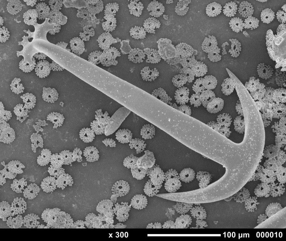
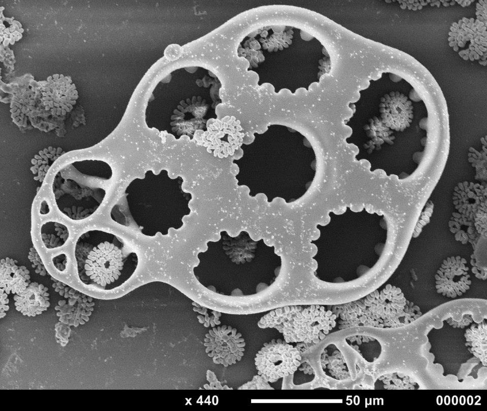
SEM was carried out to investigate finely the structures of the animal’s skin, buccal tentacles and calcareous spicules. The skin and tentacle samples were firstly fixed in pfd for 24 hours and then transferred to a 70% ethanol solution. A second sample of the skin was put in bleach, so that the skin would dissolve and the calcareous spicules would remain.
The samples were then dried via critical point drying and then mounted on the specimen stubs. The mounted samples were then sputter-coated with gold and analysed using the SEM under normal operating conditions.
Results
Image Output:
- Skin

- Tentacle

- Spicules


Acknowledgments
The author would like to thank Kathryn Green for all her help in conducting SEM on the samples. The author would also like to thank Bernie Degnan for help in collecting the samples.
|